
- Number 376 |
- November 19, 2012
New single-shot X-ray technique makes magnetic image
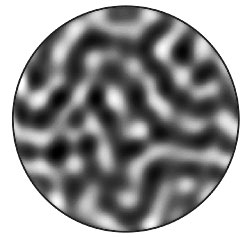
An image of a nanoscale ferromagnetic
structure made using a new single-shot
X-ray holography technique perfected at
SLAC's Linac Coherent Light Source. In the
black areas, the magnetization is "up"; in the
white areas, it's "down." The light and dark
regions are about 100 to 150 nanometers
wide. (Image courtesy Tianhan Wang)
Scientists working at SLAC National Accelerator Laboratory have captured the first single-shot X-ray microscope image of a magnetic nanostructure and shown it can be done without damaging the material.
This result not only demonstrates the success of a powerful new X-ray laser technique, but it also means that in the future researchers should be able to make movies showing tiny magnetic domains in the act of switching polarity, the process at the heart of computer hard-disk drives and future magnetic memory technologies. Understanding the details of magnetic switching could lead to faster, denser and more energy-efficient data storage devices.
The research involved 42 scientists from 16 institutions in five countries and is described in a paper in Physical Review Letters. Experiments took place at SLAC’s Linac Coherent Light Source, the world’s first hard X-ray laser, whose ultrabright, ultrashort X-ray pulses reveal never-before-seen structures and properties in matter.
Scientists aimed the X-rays at a sample made of 40 alternating layers of the metals cobalt and palladium, each layer less than a nanometer thick. Before hitting the sample, the X-rays were circularly polarized – that is, converted to waves that look like corkscrews, rotating to the left or right around the beam.
Because circularly polarized X-rays are absorbed differently by magnetic regions that are oriented “up” or “down,” they have been used for many years to image the magnetic structure of materials on the nanometer scale.
The question facing the scientists was whether they could achieve a delicate balance: single X-ray pulses strong enough to create a detailed image, yet not so energetic that they would alter the sample’s magnetic structure. If it worked, it should be possible in the future to compile single-shot images into a sequence showing magnetic changes within a sample.
The answer was an unqualified yes.
In their experiments, the scientists varied the length and intensity of the X-ray pulses. They found that longer pulses heated the sample and changed its magnetic structure. But they got good images with no damage to the sample from shorter pulses – 80 femtoseconds, or quadrillionths of a second, long – with energy fluence of 5-25 millijoules per square centimeter.
“We were delighted to find such a big window of acceptable energy density,” said Tianhan Wang, a SLAC graduate student and member of the Stanford Institute for Materials and Energy Sciences, which is run jointly by SLAC and Stanford. “Our results confirm that we should be able to make sequential, ultrafast X-ray images of a single sample.”
Wang anticipates that future research will include a detailed examination of how visible laser light can switch magnetic domains, which could lay the foundation for optically switched magnetic hard-disk drives. Today’s disk drives use electrically generated magnetic fields to write and erase data.
“To increase data density, the bits on a magnetic hard disk have to be made smaller,” Wang said. “But magnetic bits are already so small that they’re nearly unstable. More stable magnetic materials would be much more difficult to erase and rewrite data, which is undesirable.”
While laser light can be used to switch some of these stable materials, “we don’t know enough details to be able to control the process,” Wang said. “This new X-ray technique should enable us to make the measurements we need to gain such insight.”
Former SIMES researcher Andreas Scherz, now a scientist at the European X-Ray Free-Electron Laser in Germany, was the principal investigator for the study.
[Andy Freeberg, 650.926.4359,
afreeberg@SLAC.Stanford.edu]