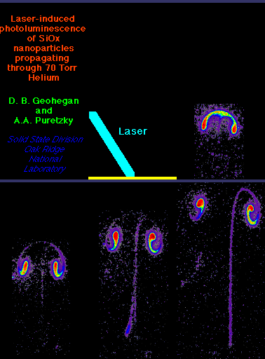
Laser ablation into a background gas creates
a bright plasma which can be detected (with sensitive ICCD-imaging) to
perhaps a few milliseconds. To induce the photoluminescence, a second
laser pulse (25 ns long, UV, 308 nm) sheet was introduced after a variable
time delay. The particles luminesced for 1.8 microseconds after this
excitation, and this light was imaged by ICCD-photography to provide a
spatial map of the nanoparticle cloud. Alternatively, we collected
the directly-scattered 308-nm light from the
nanoparticles (Rayleigh scattering, intensity proportional to nanoparticle
diameter to the sixth power) to track non-luminescing particles as well.
We used these images to understand when and where nanoparticles grow, what
causes the photoluminescence, how it can be maximized, and where the nanoparticles
go after they are formed. These first time-resolved PL
measurements from nanoparticles suspended in the gas phase were used to
maximize the luminescence of the nanoparticles in the gas phase, prior
to their deposition as photoluminescent thin films.
The background gas mass has a major effect on the
plume dynamics. Ablation of Si (m=28) into 1 Torr of (heavier,
m=40) Ar results in a uniform, stationary plume of nanoparticles while
Si ablation into lighter He (m=4) results in a turbulent ring of particles
which propagates forward at 10 m/s.[1] The graphic shows a
side-view of photoluminescent nanoparticles propagating up to 10 cm away
from a silicon target at time delays of 4 to 10 ms after the initial ablation
event.
Using these diagnostics, individual nanoparticles
which were unambiguously formed in the gas phase were collected on transmission
electron microscope grids for particle size and composition analysis using
Z-contrast imaging and electron energy loss spectroscopy. The
effects of gas flow on nanoparticle formation, photoluminescence, and collection
are described in [1].
Time-resolved photoluminescence (PL) spectra are reported for gas-suspended 1-10 nm diameter SiOx particles formed by laser ablation of Si into 1-10 Torr He and Ar. Three spectral bands (1.8, 2.5 and 3.2 eV) similar to PL from oxidized porous silicon were measured, but with a pronounced vibronic structure. Maximized violet (3.2 eV) PL from the gas-suspended nanoparticles was correlated with an ex situ SiOx (x=1.4) overall particle stoichiometry. Cryogenically-collected gas-suspended nanoparticles produced weblike-aggregate films exhibiting very weak PL. Standard anneals restored strong PL bands without vibronic structure, but otherwise in agreement with the PL measured from the gas-suspended nanoparticles.[2]
This research was sponsored by the Oak Ridge National Laboratory,
managed by Lockheed Martin Energy Research Corp., for the U.S. Department
of Energy, under contract DE-AC05-96OR22464.
2. "Photoluminescence from Gas-Suspended SiOx Nanoparticles Synthesized
by Laser Ablation" D.B. Geohegan, A.A. Puretzky, G. Duscher, and S.J.
Pennycook, Appl.Phys. Lett.73, 438 (1998) . Download
PDF file (429k)
