Research
Highlights...
 
 
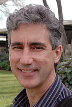
ORNL's Jerry Tuskan

ORNL's Jerry Tuskan

See below


 Number 266

July 28, 2008 

Nuclear pairs

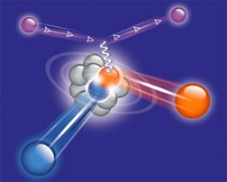
An electron interacts with a proton in a short-range correlation. The struck proton and its partner fly out of the nucleus. Graphic: Amma Shneor

An electron interacts with a proton in a short-range correlation. The struck proton and its partner fly out of the nucleus. Graphic: Amma Shneor

Like children playing a game of tag, some protons and neutrons also link up briefly inside the nucleus and then rapidly split apart. These ‘short-range correlations’ have now been quantified in the first simultaneous measurement of such pairings and their constituents. The research, conducted at DOE’s Jefferson Lab and published in the June 13 issue of Science, showed that 18 percent of all protons in the nucleus were paired with neutrons. Another one percent of protons were paired with protons, with about the same percentage of neutron/neutron pairs. When combined with theoretical calculations, the result also indicates that these pairs may have a disproportionately large effect on neutron star structure.

[Kandice Carter, 757/269-7263,
kcarter@jlab.org]

New energy storage facility under way

DOE's Sandia Labs is supporting the design of a compressed air energy storage (CAES) plant planned near Des Moines, Iowa. The Iowa Stored Energy Park (ISEP) will implement a 30-year-old, little-used technology for energy storage; only two CAES plants currently exist. CAES plants store compressed air in an underground geologic formation — in this case, an aquifer. When electricity is needed, the precompressed air is used in combustion turbines to generate electricity. While fossil fuels run the turbines, the process is more efficient. Sandia is analyzing core samples from the potential site and providing fundamental information for the design. ISEP is expected to be operational by 2012.

[Julie Hall, 505/284-7761,
jchall@sandia.gov]

A supra new type of froth

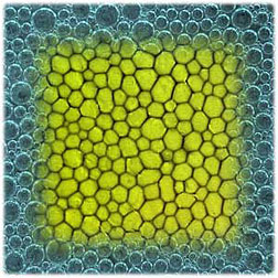
The bubble-like arrangement of magnetic domains in superconducting lead exhibits patterns very similar to conventional froths like soap foam.

A team of physicists at DOE's Ames Laboratory has found that the bubble-like arrangement of magnetic domains in superconducting lead exhibits patterns that are very similar to everyday froths like soap foam or frothed milk on a cappuccino—establishing “suprafroths” as a model system for the study of all froths. “In physics, if you can find model systems, like suprafroths, that have similar patterns, then by studying these model systems you can actually get additional information about the behavior of very complex systems like galaxies, geophysics or biophysics” says Ruslan Prozorov, the Ames Lab physicist who discovered and helped characterize suprafroths.

[Breehan Gerleman Lucchesi, 515/294-9750, breehan@ameslab.gov]

New technique a major advance for ITER

A Princeton Plasma Physics Laboratory/MIT collaboration has resulted in the demonstration of a greatly improved X-ray crystal spectrometer as a diagnostic device for ITER and other fusion reactors. ITER, which will begin operation in 2018 in Cadarache, France, is a large international fusion experiment aimed at demonstrating the scientific and technological feasibility of fusion energy. Experiments conducted recently by the PPPL/MIT team on the Alcator C-Mod fusion device at MIT mark the beginning of a new era in the ability of X-ray crystal spectrometers to measure the temperature profiles and rotational velocity of high-temperature plasmas used as the fuel for the production of fusion energy. The team’s success will benefit substantially ITER and other advanced fusion energy systems.

[Anthony DeMeo, 609/243-2755,
ademeo@pppl.gov]

Cleanup microbes enhance hydrocarbon recovery

A unique microbial consortia developed and deployed by DOE's Savannah River National Laboratory for bioremediation of oil-contaminated soils has recently been shown to be highly effective for increasing hydrocarbon recovery from oil sands without added chemicals. It may also have potential for other oil recovery initiatives, including recovery from oil shale. BioTiger(TM), which resulted from over eight years of extensive microbiology screening and characterization of samples collected from a century-old Polish waste lagoon, cleans up soils contaminated with petroleum hydrocarbons and heavy metals, and can be used to clean up petroleum spills on surfaces such as concrete bridges and building foundations. Oil sands are mined and processed to generate oil similar to that pumped from conventional oil wells, but extracting oil from these sands is more complex than conventional oil recovery. An enhanced ex situ oil recovery process using BioTiger(TM) to optimize bitumen separation could provide a means to maximize capacity and minimize environmental impact, while remaining cost effective. In a hot water extraction test using oil sands from Ft. McMurray, Canada, BioTiger(TM) demonstrated a 50% improvement in separation over 4 hours, and a five-fold increase at 25 hour.

[Angeline French, 803/725-2854, angeline.french@srnl.doe.gov]


ORNL's Tuskan seeks
secrets of trees

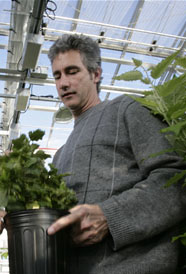
Jerry Tuskan at BioEnergy Science Center (BESC)

Jerry Tuskan says that his lifelong appreciation for trees began in the upper peninsula of Michigan and has been a constant influence, taking him down a path to a very exciting research career focused on the genetics of tree growth and development.

Tuskan came to DOE's Oak Ridge National Laboratory in 1990 to work on an early biomass feedstock program investigating alternative fuels. Coincidentally, the Human Genome Initiative happened to wrap up at about the same time as his program was ending and Tuskan saw an opportunity in the slowing sequencing facilities that had been devoted to sequencing the human genome. He submitted a proposal to sequence the first tree genome, and is now using that sequence to improve biomass production and carbon sequestration.

Accomplishing the intricate job of sequencing the massive Populus genome, which has 1.5 times more genes than the human genome, required the concerted efforts of more than 80 collaborators. "It is the most complex genome sequenced in the world. The Populus genome has undergone a duplication event, so assembling the sequence was like putting together a jigsaw puzzle of a tree reflected in a lake; you have to figure out whether the piece of the sequence belongs in the reflection or in the original,” says Tuskan.

A plant can process carbon in several different ways, incorporating it into cell walls or using it for energy production and releasing it back into the environment. Identifying the genes governing these pathways can help researchers maximize the amount of carbon that is stored stably and for long periods of time in the form of cell walls, making increasingly energy-dense biomass and removing carbon from the atmosphere.

According to Tuskan, these are heady times for studying tree growth and development: “The truth is, we really don’t know what makes a tree a tree, but we’re getting closer and closer.”—Sarah Wright

Submitted by DOE's Oak Ridge National Laboratory


Check out the joint Fermilab/SLAC publication symmetry.


DOE Pulse highlights work being done at the Department of Energy's national laboratories. DOE's laboratories house world-class facilities where more than 30,000 scientists and engineers perform cutting-edge research spanning DOE's science, energy, national security and environmental quality missions. DOE Pulse is distributed every two weeks. For more information, please contact Jeff Sherwood (jeff.sherwood
@hq.doe.gov
, 202-586-5806)

Easy to read gene chips could revolutionize medical diagnostics

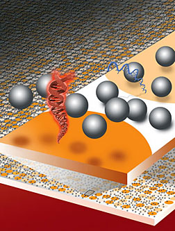
The dream of personalized medicine—in which diagnostics and treatment decisions are based on a patient’s genetic profile— may soon be expanded beyond the wealthiest of nations with state-of-the-art clinics. Berkeley Lab researchers have invented a technique in which DNA or RNA microarrays, often referred to as “gene chips,” can be read and evaluated without the need of elaborate chemical labeling or sophisticated instrumentation. Based on electrostatic repulsion—objects with the same electrical charge repel one another—the technique is relatively simple and inexpensive to implement, and can be carried out in a matter of minutes.

Berkeley Lab researchers have developed a new method for reading gene chips based on measuring the electrostatic repulsion between glass microbeads and hybridized DNA. (Illustration by Flavio Robles, Berkeley Lab Public Affairs)

“One of the most amazing things about our electrostatic detection method is that it requires nothing more than the naked eye to read out results that currently require chemical labeling and confocal laser scanners,” said Jay Groves, a chemist with Berkeley Lab’s Physical Biosciences Division, who led this research. “We believe this technique could revolutionize the use of DNA microarrays for both research and diagnostics.”

Gene chips have become one of the most powerful tools for gene-expression profiling and the identification of mutations, and for diagnostics in patients afflicted either by multiple diseases or drug-resistant strains of diseases. Until now, however, gene chip use has been limited because current techniques for reading them depend upon fluorescence detection, a demanding methodology that requires time-consuming chemical labeling, high-power excitation sources and sophisticated instrumentation for scanning.

Groves and his colleagues have developed a technique in which a fluid of electrically-charged microscopic glass beads is dispersed across the surface of a DNA microarray. Observing the Brownian motion of the beads provides measurements of the electrical charges of the DNA molecules that can be used to interrogate millions of DNA sequences at a time. What’s more, these measurements can be observed and recorded with a simple hand-held imaging device—even a cell phone camera will do.

Submitted by DOE's Lawrence Berkeley
National Laboratory

| DOE Pulse Home | Search | Comments |