- Number 302 |
- December 21, 2009
A speedy CAT scan for cells
Three-dimensional tomography
of Candida albicans
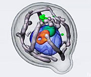
A team from DOE’s Lawrence Berkeley National Laboratory, Stanford University, and the University of California at Berkeley have used the National Center for X-ray Tomography at Berkeley Lab’s Advanced Light Source (ALS) to make 3-D movies of the internal structure of Candida albicans, a microbe that can cause yeast infections, capturing the changes that occur when the organism is exposed to a new and promising antifungal therapy. The soft X-ray tomography images revealed that microbes treated with the candidate drug did not develop the characteristic structures that make C. albicans infectious. Tracking the changes would have taken months of work using other microscopy techniques, and a few would have been missed altogether.
“We saw cellular changes that hadn’t been seen in the past,” says Gerry McDermott of Berkeley Lab’s Physical Biosciences Division and the University of California at San Francisco. “Because of its high resolution and very little sample prep time, this new technology may help expedite drug discovery.”
The ALS’s National Center for X-ray Tomography is the only soft X-ray tomography facility dedicated to biochemical and biomedical research.[Paul Preuss, 510.496.6249,
paul_preuss@lbl.gov]