
- Number 335 |
- April 18, 2011
Modeling radiation energy deposition in a complex biological system
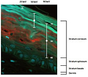
Using a microscope,
scientists looked at a
vertical slice through a
skin-tissue model showing
how the radiation
penetrated the skin. The
centers of the bars mark
the depths at which half of
the electrons are expected
to stop.
Using new tools developed at DOE’s Pacific Northwest National Laboratory, scientists are seeing how the skin responds to low doses of radiation, a hazard in certain medical procedures and jobs. The scientists at PNNL and Washington State University-Tri Cities are modeling the data from these experiments to understand the mechanisms involved when cells encounter low levels of radiation.
In this study, the WSU-TC and PNNL scientists used an artificial skin model made of normal human epidermal skin cells and connective tissue cells called fibroblasts. This model has a well-defined cellular composition that scientists can modify to see how different cell types interact after irradiation. It also provides a view that more closely matches the real world, not that seen in a Petri dish.
This work is helping to develop biologically based risk models and help ensure that radiation protection standards are adequate and appropriate. The Department of Energy's Low Dose Radiation Research Program funded the research.
[Kristin Manke, 509.372.6011,
kristin.manke@pnl.gov]