
- Number 351 |
- November 28, 2011
Building block detectors for plants
The Phytotron is a laboratory
built to study plants.
Plants face all kinds of stress. Bugs chew on them, they get fungal infections, and they can get too hot or cold. So how do plants respond? Researchers at DOE's Jefferson Lab are developing tools to help biologists find out.
Members of Jefferson Lab's Radiation Detector and Imaging group are developing new imaging tools to help researchers at Duke University's Phytotron find out how plants respond to stress. The Phytotron is a laboratory built to study plants. It has so-called Environmental Growth Chambers that allow researchers to control nearly every aspect of a plant's environment, from the nutrients it gets in the soil to the relative humidity and pollutants in the air. Fine-tuning individual aspects of a plant's surroundings can help researchers identify those aspects that can help or harm plants.
While a plant's overall health can often be determined through simple observation, researchers sometimes need to see what's happening on the inside. That's where Jefferson Lab group leader Drew Weisenberger and his colleagues come in. They are working to develop tools that can image inside plants.
"It's at the point where biologists are kind of learning what we can provide and we're trying to learn what they need. So they have to learn a little physics and we have to learn a little biology. We're just trying to develop the tools and trying to get the tools to work," Weisenberger says.
For instance, a study with the common barley plant addresses how it would respond to higher levels of carbon dioxide in the atmosphere. Green plants use CO2 in photosynthesis to create their own food. Researchers want to follow the path of carbon from the CO2 inside plants, from the moment it is breathed in by the plant, through its photosynthesis into sugars, to storage in the plant's roots.
"We're just coming up with tools to help them quantify the physiology of photosynthesis under different conditions and parameters," Weisenberger adds. "They are also interested in studying invasive species and understanding how photosynthesis utilization and carbon dioxide utilization is different in various species,"
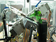
For the barley experiment, a special form of CO2 was generated at the Triangle Universities Nuclear Laboratory at Duke. The carbon atoms in this CO2 emit positrons, which are the anti-particles of electrons. When a positron encounters an electron in one of the atoms of the plant, the two particles annihilate each other and produce gamma rays. Those gamma rays can be detected by the Jefferson Lab group's PhytoPET system.
The PhytoPET system functions much like the PET scanners found in hospitals. One key difference is that the parts of the PhytoPET that collect the tell-tale gamma rays can be custom fit to the contours of a small plant. The building-block-inspired system is designed to give accurate information whether the blocks are arranged in a circular, semi-circular, flat or other configuration.
The Duke researchers also want to better understand how plants respond to injury, such as that inflicted by a bug slowly munching on it or by some type of infection. A new study with an oak tree seedling has recently been undertaken to determine whether the plants tap the sugar stored in their roots to combat infection.
"They introduce a fungus into the roots and see if that infected plant tries to deal with its infection. They didn't really have a way to measure that accurately, without destroying the plant," Weisenberger explains.
Weisenberger and his colleagues designed a system that images the thin roots of plants. The new system, called PhytoBeta, is capable of detecting fast-moving positrons directly, allowing it to capture information from thin parts of the plant that may be missed by PhytoPET.
What's more, PhytoPET and PhytoBeta can be used together to image all parts of plants, so biologists get the most information they can out of each experiment.
Weisenberger says there's still much work to be done on the two systems, including making sure they meet the biologists' needs without interfering with experiments, such as making the new detectors highly reflective.
"Inside a growth chamber, everything is aluminized. Biologists don't want you to cut off the light: They don't want to interfere with it. That's why we make our detectors reflective now, and that's kind of different from what we're used to," Weisenberger says.
The work is supported by the Department of Energy's Office of Biological and Environmental Research. It builds on the group's many other projects that go beyond imaging subatomic particles produced in experiments at Jefferson Lab, including breast imaging and small animal imaging.[Kandice Carter, 757.269.7263,
kcarter@jlab.org]