Archive Site Provided for Historical Purposes

Sponsored by the U.S. Department of Energy Human Genome Program
In this issue...
Available in PDF
In the News
Comparative Genomics
Mouse
Web, Publications, Resources
Funding
Meeting Calendars & Acronyms
Although typically focused on only one part of DOE's Biological and Environmental Research (BER), Human Genome News will now include material from the Medical Sciences Division (MSD), which shares the same mission. MSD's nuclear imaging has all but eliminated the need for exploratory surgeries.
Following is a summary of MSD's new booklet, Converting Energy to Medical Progress (April 2001). The booklet is available from HGMIS and can be downloaded from the Web (https://www.doemedicalsciences.org/).
Nuclear medicine is an exciting field in healthcare that provides important information for diagnosing, evaluating, and managing disease. Virtually all hospitals, as well as many clinics and doctors' offices, conduct nuclear medicine tests and scans. About 13 million (35,000 a day) such procedures are performed each year on patients in the United States (and many more in other countries) in cardiology, oncology, neurology, sports and internal medicine, thyroid disorders, surgery, gastrointestinal ailments, pulmonary disorders, infection, and dementia.
Nearly every nuclear medicine scan or test used today was made possible by research funded by BER and its predecessor agencies on radiotracers, radiation-detection devices, gamma cameras, positron emission tomography (PET) and single-photon emission computed tomography (SPECT) scanners, and computer science.
In managing DOE's nuclear medicine research program, MSD pursues two main areas of scientific investigation imaging systems and radiopharmaceuticals (radiotracers). The aim is to develop beneficial applications of nuclear technologies for medical diagnosis and treatment of many diseases.
Biological Imaging
All human characteristics depend on a galaxy of biochemical reactions that occur many millions of times per minute within the cells and tissues of the body. A deranged chemical process can cause disease, resulting in other abnormal biochemical (physiological) changes. With its unique ability to reveal biochemical processes, nuclear medicine provides crucial information about numerous diseases. Nuclear medicine procedures are different from X rays, scans by computed tomography (called CT) and magnetic resonance imaging (called MRI), and ultrasound, all of which primarily visualize structure and shape (anatomy).
Nuclear medicine images are produced by low levels of energy emitted from medically useful radiotracers introduced into a patient's body. SPECT gives off gamma rays and PET emits positrons, another form of energy that converts to gamma rays. Radiotracers are designed to provide insights about healthy, normal biology, the biological process of disease, and even the molecular errors that cause disease.
Radiotracers interact with such biological processes as bone mineral turnover, potassium transport in heart muscle, or glucose metabolism in various organs or tumors. Highly sensitive scanners detect and process the energy signals, after which computer programs reconstruct them into diagnostic images. PET and SPECT, for example, produce 3-D images that look like multiple slices through the body.
Imaging Gene Expression
BER scientists have successfully created images of genetically altered organ function in animals. Now, MSD has initiated exploratory research to develop new radiotracers based on messenger RNA for dynamic imaging of gene expression in animals in real time. BER researchers at Sloan-Kettering, for example, created iodine-124 FAIU, a highly specific radiopharmaceutical that provides the first nuclear medicine images showing the expression of certain genes in tumors in a live animal.
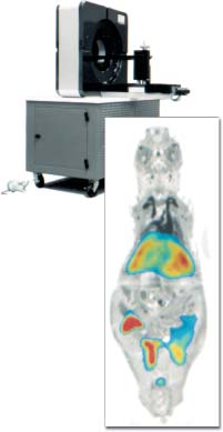
Miniature PET scanner, the "microPET" for imaging mice, developed at the University of California, Los Angeles, with scan inset.
As scientists discover more information about the relationship between genes and disease or behavior, they can identify new molecular targets for imaging the biological activity of disease. In time, drugs may be custom made for individual patients based on genetic "fingerprinting," and nuclear medicine will play a crucial role in this pursuit.
PET imaging techniques developed at Washington University, for example, are helping to identify which patients with breast cancer will respond to tamoxifen hormone therapy. Scientists there also have developed fluorine-18 fluoroestradiol that targets estrogen receptors on breast tumors. The presence or absence of abundant estrogen receptors in breast cancer cells can help doctors select the most appropriate chemotherapy for these patients.
Since mice can be engineered biologically to carry genes that produce disease, molecular probes such as microPET allow the imaging of disease initiation and progression in a living mouse. In concert with this research, scientists are investigating highly sophisticated drugs designed to correct the molecular errors of disease. Combined with the explosive growth of knowledge from genome research, PET and microPET play a major role in the promising new era of molecular diagnostics and therapeutics.
Future Impacts
The nuclear medicine of tomorrow will depend on the discovery of radiopharmaceuticals that seek specific molecular and genetic targets, the design of companion advanced scanners for creating meaningful images, and the promise of new radiopharmaceutical treatments for cancers and genetic diseases.
The electronic form of the newsletter may be cited in the following style:
Human Genome Program, U.S. Department of Energy, Human Genome News (v11n3-4).
The Human Genome Project (HGP) was an international 13-year effort, 1990 to 2003. Primary goals were to discover the complete set of human genes and make them accessible for further biological study, and determine the complete sequence of DNA bases in the human genome. See Timeline for more HGP history.

Published from 1989 until 2002, this newsletter facilitated HGP communication, helped prevent duplication of research effort, and informed persons interested in genome research.
