Spectroscopic and Imaging Diagnostics Facility

Experimental
Techniques Used for Time-Resolved Diagnostics of Thin Film and Nanomaterials
Growth Processes
Fast plasma diagnostics (5 ns resolution) are applied in combination to study
laser ablation plumes during materials synthesis. A 25-ns pulsed laser beam
is typically used to remove material from a target (ablation). The plume of
vaporized material is typically a partially-ionized laser plasma which emits
light. This light is imaged using gated ICCD (intensified-CCD array) photography,
or analyzed with optical absorption spectroscopy or optical emission
spectroscopy. The material is partially ionized and fast ion probes
conveniently track the flux of fast moving ions. For weak ablation (desorption),
a second (time-delayed) laser must be used to illuminate the plume to generate
laser-induced fluorescence or for small gas-suspended nanoparticles,
laser induced photoluminescence. This light is then imaged using ICCD-photography
and spectroscopically analyzed. Rayleigh scattering imaging is used
to locate and track nanoparticles or nanotubes as they form, transport, and
deposit. Gated photon counting is used to investigate the traces of
light from weakly-emitting plumes or the blackbody emission from hot particulates.
Time-of-flight mass spectrometry (TOFMS) is also used to mass-analyze
laser-desorbed targets. A reflectron TOFMS and a linear TOFMS with delayed
pulse extraction electronics and high-voltage post accelerator detectors are
employed to investigate heavy cluster ions.
Facilities Description and Example
Two setups for in situ imaging and spectroscopy of laser-ablation processes and laser-processing utilize two excimer lasers and one Nd:YAG laser, two gated-intensified CCD-arrays, gated-intensified diode array, and several spectrometers.
Two chambers are used: a general-purpose ablation chamber (turbopumped) for thin film and nanoparticle synthesis in vacuum or room-temperature ambients, and a 2" windowed tube furnace for studies of nanomaterial synthesis at high temperatures and variable pressures.
In general, one laser (either a Nd:YAG or one excimer laser) is used to ablate a target. A second laser (excimer) is time-delayed using a delay generator and used to probe the plume of material generated by the first laser. Laser-induced fluorescence, incandescence, or scattering is then imaged or collected by a spectrometer using one of the gated-intensified detectors. These detectors can be triggered to occur coincident, or slightly delayed from the probe laser. A variety of techniques are employed.
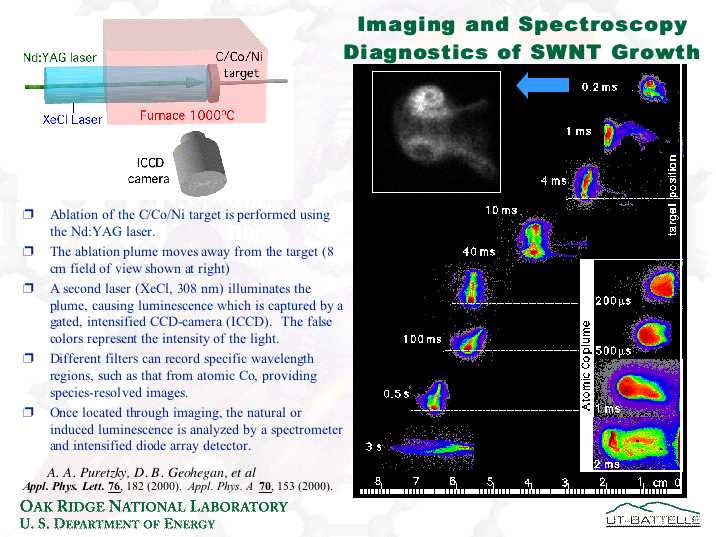
Imaging and Spectroscopy of the C/Ni/Co plume inside a tube furnace at 1000 °C
Slide 1: Imaging measurements locate the plume location. Plasma luminescence lasts only 0.2 ms. After that, the XeCl laser is needed to "light up" the plume.
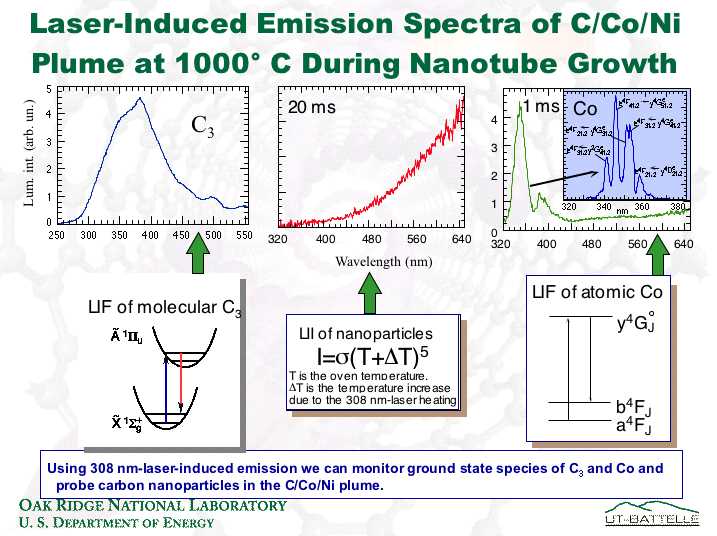
Slide 2: In addition to the bright emission from the laser plasma (during the first 0.2 ms), the 308-nm laser can excite ground-state molecules and atoms, as well as clusters.
Slide 3: A compendium of the spectra obtained for the images of slide 1. Red curves are luminescence induced by the 2nd laser, blue curves show how the natural luminescence compares (dies away by 0.2 ms, in agreement with the imaging observations).
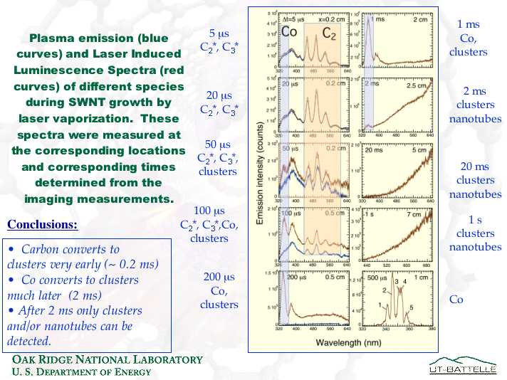
The key finding of these measurements was that the atomic and molecular species' populations appear to feed the formation of clusters within 2 ms. The majority of nanotube growth occurs for times > 2 ms (derived from separate measurements where imaging was used to limit the time spent by the plume in the hot oven). Therefore, the feedstock for the majority of nanotube growth is small carbonaceous clusters, or larger aggregates of these clusters (and not atomic or molecular species).
Back to