Carbon Nanotube Synthesis and
Diagnostics

At ORNL, we are growing single-walled
carbon nanotubes (SWNT) for use in composite materials and in molecular electronic
devices. Laser ablation is one of the best ways to produce high-quality SWNT.
Lack of information on the local
environmental conditions during the growth of carbon nanotubes has hindered
efforts to understand the formation of these unique nanomaterials. Moreover,
SWNT are currently produced in ~ gram/day quantities, apparently growing at
a very small fraction of their theoretical growth rate. If the growth process
could be understood and optimized, the incredible properties of carbon nanotubes
could be applied for a multitude of high-volume applications. We intend to understand
and optimize the growth process by developing and applying in situ diagnostics
of the nanotube growth process.
Recently we have developed a set
of unique diagnostics including spectroscopic gated-ICCD imaging, ion probe
measurements and several optical spectroscopic methods to monitor the laser
ablation of graphite for the deposition of DLC films and the synthesis of fullerenes.
We propose to extend these measurements to understand how carbon nanotubes nucleate
and grow.
Unlike continuous production of carbon
nanotubes by the arc-discharge and CVD-deposition methods, the pulsed laser
vaporization (PLV) method is amenable to time-resolved measurements of nanotube
formation and growth. This is because the plume of starting material is created
very rapidly using a short (~10 ns) laser pulse that gives well defined initial
conditions for the conversion of the starting material into clusters and for
nanotube growth. Understanding why laser ablation produces such high nanotube
yields is a high priority.
In the PLV method, a laser pulse
evaporates a solid target of graphite which contains a small amount of metal
catalyst (~1 atomic % Ni and ~1% Co) into a background gas (~500 Torr of Ar)
which is gently flowing through a quartz tube inside a high temperature (~1000
C) oven. The laser converts a small amount of the composite solid material into
a plasma of atoms and molecules which contains mainly C, C2, C3, Ni, Co and
their ions. These species leave the target with extremely high initial velocities
~(from 1-5)x106 cm/s. This material violently collides with the background gas,
generating a series of shock waves which provides additional heating that dissociates
and ionizes the initial ejecta. Basically, however, the initial ejecta are confined
by the background gas to remain inside a "bubble" of hot plasma which thermalizes
as it expands in a stepwise way. In summary, the starting material for nanotube
growth is a hot, expanding plasma of atomic/molecular carbon species and atomic
vapor of catalyst.
The composition and other parameters
of this plasma can be obtained using plasma
diagnostic techniques. After cooling and partial recombination of this confined
plasma plume the vapor condensation begins that forms clusters of carbon and
metal catalyst. Our spectroscopic diagnostics
allows us to study clustering within the laser ablation plume. These diagnostics
can provide the key information about clustering times for carbon and catalyst
in the nanotube growth environmental conditions.
Shown below are ICCD-images which
reveal the complex way in which a plume of vaporized graphite expands into room-temperature
argon gas (300 Torr) following laser-vaporization by a 46-nanosecond laser pulse.
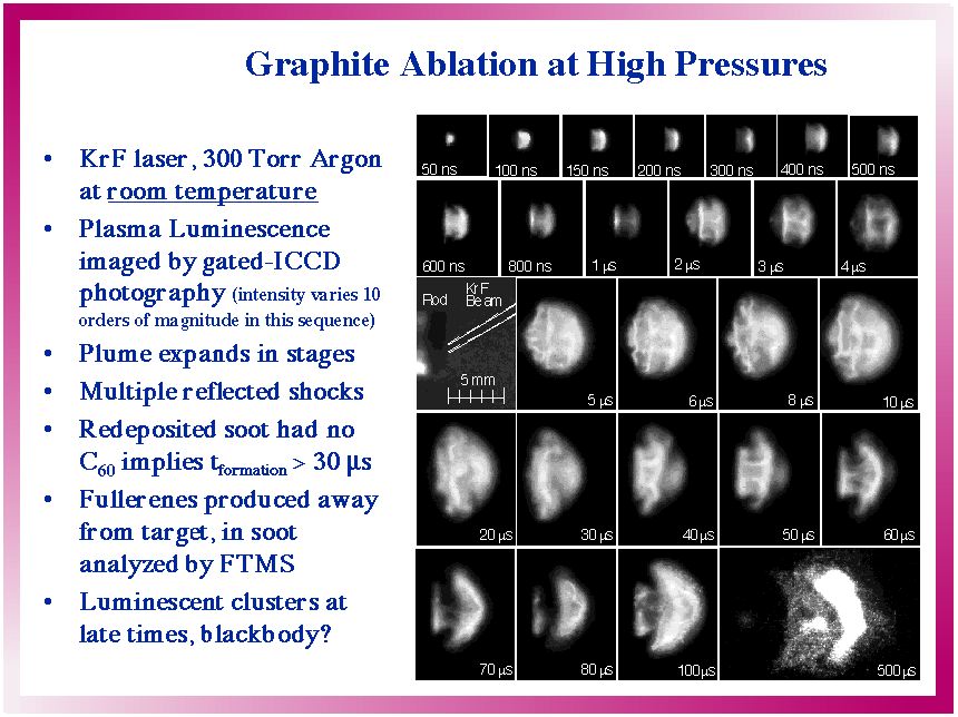
Animated, this looks like...
We are currently using optical spectroscopy to probe the species, temperature,
and size of particles in this plume at the different times ( and much longer
times than) shown in the above images. Moreover, we are studying these processes
under the exact conditions for nanotube growth inside the hot oven (as shown
at the top) to understand how carbon nanotubes grow. We are correlating the
collected nanotubes with the growth conditions characterized by the diagnostics
described below...
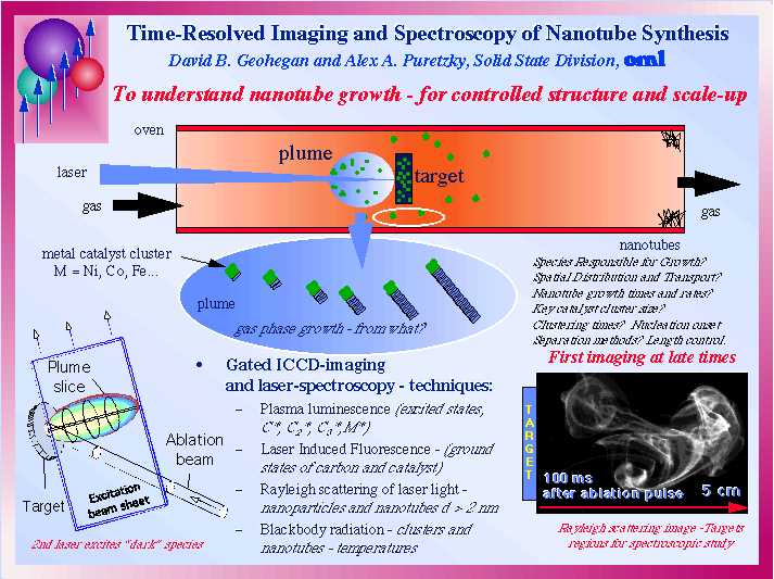
We are developing several in situ
diagnostic techniques to characterize nanotubes as they grow. Gated intensified
CCD-array photography (ICCD Photography) is useful to characterize the
initial plasma luminescence (as shown in the first slide and animation above).
Once the spatial and temporal locations of the plume have been identified via
imaging, light can be collected at these positions and optical emission spectroscopy(OES)
can be used to identify the excited states in the plume. Laser-induced
fluorescence(LIF)or optical absorption spectroscopy(OAS) are needed
to probe "dark" material in the ground states of atoms, molecules, and
small clusters. In this case, a second (probe) laser pulse (~25 ns long) is
introduced at the time-delay of interest, providing excitation to the ground-state
species to induce luminescence from excited-states. This light can be imaged
or spectroscopically-analyzed to determine populations (and lifetimes) of species
in the plume. As condensation occurs and nanoparticles or nanotubes are formed,
the probe laser light which is directly scattered from the particles can be
imaged to reveal their presence. Rayleigh scattering (RS) imaging is
effective for particles larger than just 2-10 nm, so it can be used to see nanotubes
as they are just beginning growth into micron-long nanowires. In the oven, these
nanotubes are hot and emit blackbody radiation (BB) which can also be
imaged, or spectroscopically collected and curve-fit to reveal their temperature.
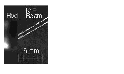
Shown above is an example of a RS-image taken at room temperature for graphite
ablation into 300 Torr Argon. These techniques, and others, are currently being
implemented inside the hot oven to understand nanotube growth at high temperatures.
A compendium of our results is shown
below, which shows actual (false-color) images of the ablation plume vs. time
(log scale), the species detected spectroscopically from the plasma emission
or laser-induced luminescence spectra, plume temperatures measured through blackbody
emission above the oven temperature, and lengths measured from arrested-growth
experiments.
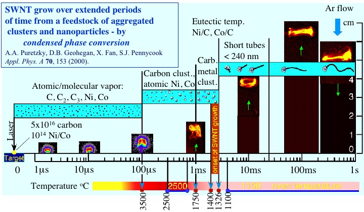
The data shows that carbon in the
plume condenses within the first 0.2 ms after laser vaporization. At this point,
the plume changes appearance, as the clusters begin to aggregate and become
trapped in a swirling vortex ring (smoke ring). However, the atomic Co and Ni
in the plume condense at lower temperatures, and wait for the plume temperature
to cool. The ground-state Co population, for example, peaks at 1 ms as excited
atoms relax into their ground states. Over the next millisecond, the ground
state metal atoms condense, such that by t = 2 ms, the plume is virtually entirely
composed of clusters, trapped within the swirling smoke ring. This "microreactor"
is where the single-walled carbon nanotubes grow. If growth is stopped after
20 ms, only short SWNT are found (around 200 nm long), indicating that the majority
of their growth (to 10 micron lengths) occurs over long times – 100 ms
to seconds of time – at rates of 0.5–5 microns/second. The feedstock
for growth at these long times are the condensed cluster aggregates which remain
in close proximity to the growing nanotubes thanks to the vortex dynamics of
the swirling "smoke ring".
References available on the growth
of SWNT by laser vaporization probed using in situ spectroscopic and imaging
diagnostics
Investigations of single-wall carbon nanotube growth by time-restricted
laser vaporization
Alex A. Puretzky, Henrik Schittenhelm, Xudong Fan, Michael J. Lance, Larry
F. Allard, Jr., and David B. Geohegan
Phys. Rev. B 65, 245425 (2002) Download
PDF file (1000 KB)
"Dynamics of single-wall carbon nanotube synthesis by laser vaporization"
A.A. Puretzky, D.B. Geohegan, X. Fan, S.J. Pennycook
Appl. Phys. A 70, 153, (2000).
Download PDF file (531 KB)
In situ imaging and spectroscopy of single-wall carbon nanotube synthesis
by laser vaporization
A. A. Puretzky, D. B. Geohegan, X. Fan, and S. J. Pennycook,
Appl. Phys. Lett. 76, 182, (2000). Download
PDF file (304 KB)
Images in first slide above published
in...
"Time Resolved Diagnostics of Excimer Laser Generated Ablation Plasmas Used
For Pulsed Laser Deposition", David B. Geohegan, p. 165 inExcimer Lasers:
The Tools, Fundamental Processes and Applications, , Kluwer Academic Publishers,
Netherlands, (1994).
"Gated ICCD Photography of the KrF-Laser Ablation of Graphite into Background
Gases," David B. Geohegan, A. A. Puretzky, R. L. Hettich, X.-Y. Zheng, R. E.
Haufler, and R. N. Compton, in Advanced Materials '93, IV/ Laser and Ion
Beam Modification of Materials, edited by I. Yamada, et al. IUMRS-ICAM Conference
, Trans. Mat. Res. Soc. Jpn., 17, 349 (1994).
see also
"Laser Ablation of Graphite in Different Buffer Gases" A. A. Puretzky, D. B.
Geohegan, R. E. Haufler, R. L. Hettich, , X.-Y. Zheng, and R. N. Compton, p.
365 inLaser Ablation: Mechanisms and Applications II, edited by J. C.
Miller and D. B. Geohegan, Amer. Inst. of Physics Conf. Proc. 288 , Amer.
Inst. of Physics, New York , (1994 ).
