
- Number 382 |
- February 18, 2013
New look at cell membrane reveals surprising organization
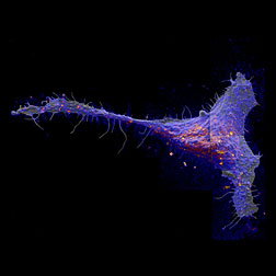
The local abundance of metabolically
incorporated 15N-sphingolipids in the plasma
membrane of a fibroblast cell, overlaid on
the corresponding secondary electron image.
Red and yellow colors are local elevations
in sphingolipid abundance.
Image by Kevin Carpenter/LLNL.
A new way of looking at a cell's surface reveals the distribution of small molecules in the cell membrane, changing the understanding of its organization.
A novel imaging study by researchers fromDOE's Lawrence Livermore National Laboratory, the University of Illinois and the National Institutes of Health revealed some unexpected relationships among molecules within cell membranes.
Their findings provide a new way of studying cell structure and ultimately its function.
Led by Mary Kraft of the University of Illinois, Peter Weber of Lawrence Livermore National Laboratory and Joshua Zimmerberg of the National Institutes of Health, the team published their findings in the online version of the Jan. 28 edition of the Proceedings of the National Academy of Sciences.
Cells are enveloped in a semi-permeable membrane that acts as a barrier between the inside and outside of the cell. The membrane is mainly composed of a class of molecules called lipids, which are small and easily perturbed when tracked.
"Lipids have multiple functions serving as both membrane structure and signaling molecules, so they regulate other functions inside the cell," Kraft said. "Therefore, understanding how they're organized is important. You need to know where they are to figure out how they're performing these regulatory functions."
Previous cell membrane research suggested that lipids in the membrane assemble into patches, called domains, which differ in composition. But the challenge of direct observation has limited research into how lipids are organized in the membrane, and how that organization affects cell function. In the new study, the team used an advanced, molecule-specific imaging method developed at Lawrence Livermore that allowed the researchers to look at the membrane itself and map a particular type of lipid on mouse cell membranes. The researchers at University of Illinois fed lipids labeled with rare stable isotopes to the cells and then imaged the distribution of the isotopes with high-resolution imaging mass spectrometry at LLNL.
Called sphingolipids (sfing-go-lipids), these molecules are thought to associate with cholesterol to form small domains about 200 nanometers across. The direct imaging method revealed that sphingolipids do form domains, but not in the way the researchers expected.[Anne M. Stark, 925.422.9799,
Stark8@llnl.gov]